Up
Leaf fibers
Drimys wood
Oak wood
Flax fibers
Vessels
Pits, xs
Wood f., ls
Pine pits
Dicot pits
Monocot bundles
Living fibers
Dead fibers
Stone cells
Stone c., mag
Stone c., polarized
Macrosclereids
Macro., young
Sweet olive
Astrosclereid
Astro., mag
Astro., hi mag
Astro., body
Astro., arms
Libriform fibers
Phloem fibers
Maceration
Fiber-tracheid
Fiber bundle
F. bundles, mag
Leaf margin
Epidermis
Gelatinous f.
| |
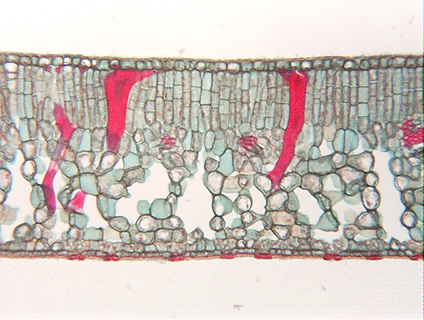 Fig. 5.3-6.
Transverse section through leaf of sweet olive (Osmanthus). The macrosclereids
(red) in this leaf occur as idioblasts, that is,
each is surrounded by cells that are not like itself. Consequently, their shape
is easily visible. The small brown streaks and splotches in these are the
remnants of the lumen, all the rest of the macrosclereid is secondary wall (each
has a primary wall too thin to be seen here). These macrosclereids are so large,
and are lying in varied directions, so as the microtome knife cut through the
leaf, it often cut off one or both ends of each sclereid. Fig. 5.3-6.
Transverse section through leaf of sweet olive (Osmanthus). The macrosclereids
(red) in this leaf occur as idioblasts, that is,
each is surrounded by cells that are not like itself. Consequently, their shape
is easily visible. The small brown streaks and splotches in these are the
remnants of the lumen, all the rest of the macrosclereid is secondary wall (each
has a primary wall too thin to be seen here). These macrosclereids are so large,
and are lying in varied directions, so as the microtome knife cut through the
leaf, it often cut off one or both ends of each sclereid.
These sclereids cannot provide strength to the leaf since they occur as
idioblasts rather than as an extensive mass. They probably deter insects from
eating the leaf – each bite would encounter some tough, difficult to chew,
non-nutritious sclereids.
|