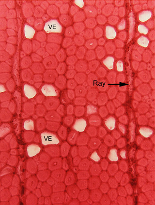
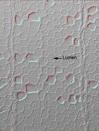
Fig. 5.1-3a and b. Transverse section of wood of oak (Quercus). Most of the cells here (the ones that are almost solid red) are xylary fibers, cut in transverse section, so they appear round rather than long. They are so red because the cell is little more than just secondary wall: the primary walls are the fine dark lines that form the outline of the cells, and the tiny dot in the center of each cell is the lumen, the space where the protoplasm had been located. The primary walls and lumens stand out a bit more in "b" which was modified on Adobe Photoshop to accentuate edges. When the cells began depositing their secondary wall, the lumen and protoplast filled all the space from the cell’s primary wall on one side to its primary wall on the other. But as the cell deposited secondary wall along the inner surface of the primary wall, the protoplast was forced to shrink. As the secondary wall continued to thicken, the protoplast had to continue to shrink, probably mostly by allowing the central vacuole to shrink. At some point, there would have been almost no vacuole, very few other organelles, and the nucleus would have been squeezed into a narrow, long, threadlike shape. Despite this, the cell would have continued synthesizing cellulose, hemicelluloses, lignin and other components of the secondary wall. Finally, the cell would have died.
Some cells have a large white lumen and relatively thin red walls – these are vessel elements, sclerenchyma cells that conduct water. Although their secondary walls are not nearly as thick as those of the fibers, they are substantial nonetheless. Also, notice that in some fibers, there is a bit of white lumen present, in others the lumen is just a tiny dark dot. Fibers are tapered cells, wider in the middle and narrower at the ends, and the lumen has the same shape: those fibers with a bit of white lumen showing have been cut near their middle region, those with just a dot were cut near one of their ends.
The tall narrow cells are ray cells.