 Fig.
5.1-4a and b.
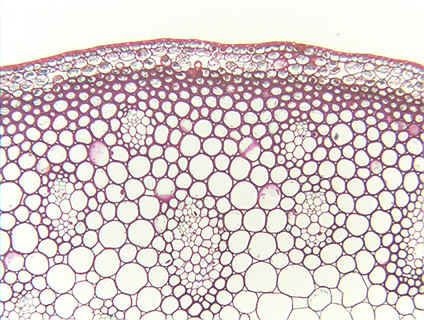
Transverse section of stem of flax (Linum). Although these cells have
thick secondary walls, they are such broad cells that the walls appear
comparatively thin. Two features indicate they are
sclerenchyma, however: 1) they are stained red, and 2) the presence
of pits, the fine white gaps in the secondary walls (arrows in lower
micrograph). In almost all cases here,
you can see that the pits occur as pit-pairs: a pit in the secondary wall of one
cell faces a corresponding pit in the secondary wall of the adjacent cell. Look
carefully and you will see that there is always a pit-membrane
between the two pits of a pit-pair: pits are not holes all the way from one cell
to another, they are just areas where there is no secondary wall (but the two
primary walls and middle lamella are still present; see Fig. 5.5 in Plant
Anatomy.).
Fig.
5.1-4a and b.
Transverse section of stem of flax (Linum). Although these cells have
thick secondary walls, they are such broad cells that the walls appear
comparatively thin. Two features indicate they are
sclerenchyma, however: 1) they are stained red, and 2) the presence
of pits, the fine white gaps in the secondary walls (arrows in lower
micrograph). In almost all cases here,
you can see that the pits occur as pit-pairs: a pit in the secondary wall of one
cell faces a corresponding pit in the secondary wall of the adjacent cell. Look
carefully and you will see that there is always a pit-membrane
between the two pits of a pit-pair: pits are not holes all the way from one cell
to another, they are just areas where there is no secondary wall (but the two
primary walls and middle lamella are still present; see Fig. 5.5 in Plant
Anatomy.).
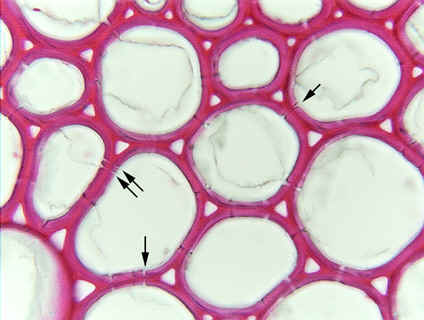 Pits
are typically extremely narrow, usually only 1 to 2mm
in diameter; even though the lower micrograph was taken at high magnification, the
details of the pits and membranes are difficult to see. However, you can see
that the pits are basically narrow holes with straight sides – they do not
curve or have one part wider than the other. Consequently, these are simple
pits.
Pits
are typically extremely narrow, usually only 1 to 2mm
in diameter; even though the lower micrograph was taken at high magnification, the
details of the pits and membranes are difficult to see. However, you can see
that the pits are basically narrow holes with straight sides – they do not
curve or have one part wider than the other. Consequently, these are simple
pits.