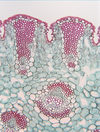
 Fig. 5.3-16.
Transverse section of leaf of yucca (Yucca). Yuccas have large, thick
leaves that have a tough, leathery texture due to having many bundles of fiber
cells. The fiber bundles are recognizable in
the high magnification view (below, at the bottom of this page) as consisting of hundreds of fibers located just
interior to the epidermis and hypodermis. Because these fibers are not part of
the xylem, they are extraxylary fibers. Each of the two big masses of fibers has
a lighter region along its inner edge; that region is a bit of phloem and xylem.
Because the bundle has both vascular tissue and many fibers, it is a fibrovascular
bundle. At the center of the micrograph is another fibrovascular
bundle in which the vascular tissue is more abundant: the red cells in the
center are xylem conducting cells, the very tiny green cells above the xylem are
phloem cells, and above and below the conducting cells are two arc of red fiber
cells.
Fig. 5.3-16.
Transverse section of leaf of yucca (Yucca). Yuccas have large, thick
leaves that have a tough, leathery texture due to having many bundles of fiber
cells. The fiber bundles are recognizable in
the high magnification view (below, at the bottom of this page) as consisting of hundreds of fibers located just
interior to the epidermis and hypodermis. Because these fibers are not part of
the xylem, they are extraxylary fibers. Each of the two big masses of fibers has
a lighter region along its inner edge; that region is a bit of phloem and xylem.
Because the bundle has both vascular tissue and many fibers, it is a fibrovascular
bundle. At the center of the micrograph is another fibrovascular
bundle in which the vascular tissue is more abundant: the red cells in the
center are xylem conducting cells, the very tiny green cells above the xylem are
phloem cells, and above and below the conducting cells are two arc of red fiber
cells.
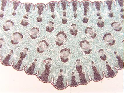
 The low magnification view shows that
fibrovascular bundles constitute a large fraction of the leaf’s volume, with
the fiber-rich type of fibrovascular bundle being abundant along the leaf’s
surface, the conducing cell-rich type of bundle being restricted to the interior
of the leaf.
The low magnification view shows that
fibrovascular bundles constitute a large fraction of the leaf’s volume, with
the fiber-rich type of fibrovascular bundle being abundant along the leaf’s
surface, the conducing cell-rich type of bundle being restricted to the interior
of the leaf.