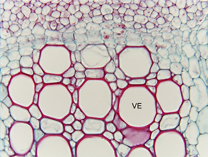
 Fig.
5.1-5. Transverse section of ragweed stem (Ambrosia). These
cells resemble the fibers of flax (Fig. 5.1-4), but these are vessel elements
instead, involved in water conduction rather than in providing elastic strength.
In transverse sections, it can be difficult to be certain if a cell is a vessel
element or a fiber; features to consider: 1)
very large cells (more than 30mm
in diameter) with open lumens are almost certainly vessels, fibers are almost
never so wide. 2) Fibers almost always occur as masses of fibers – most fibers
have neighbors that are other fibers, so the cells here – each surrounded by
thin-walled parenchyma cells with protoplasts – do not fit this criterion for
fibers. In wood, vessel elements are often also completely surrounded by
sclerenchyma (other vessels and fibers) and then it can be difficult to
distinguish between fibers and narrow vessels. 3) Fibers will have narrow pits
that are usually not very abundant; it might be necessary to look at a number of
sections before actually seeing a pit (the flax fibers in Fig. 5.1-4 have an
unusually high density of pits). Vessel elements on the other hand have a high
density of very wide pits that have a border on their inner aperture.
Unfortunately, to see these bordered pits in transverse sections, it is
necessary to have very good sections and a good microscope; see Fig. 5.1-6.
Fig.
5.1-5. Transverse section of ragweed stem (Ambrosia). These
cells resemble the fibers of flax (Fig. 5.1-4), but these are vessel elements
instead, involved in water conduction rather than in providing elastic strength.
In transverse sections, it can be difficult to be certain if a cell is a vessel
element or a fiber; features to consider: 1)
very large cells (more than 30mm
in diameter) with open lumens are almost certainly vessels, fibers are almost
never so wide. 2) Fibers almost always occur as masses of fibers – most fibers
have neighbors that are other fibers, so the cells here – each surrounded by
thin-walled parenchyma cells with protoplasts – do not fit this criterion for
fibers. In wood, vessel elements are often also completely surrounded by
sclerenchyma (other vessels and fibers) and then it can be difficult to
distinguish between fibers and narrow vessels. 3) Fibers will have narrow pits
that are usually not very abundant; it might be necessary to look at a number of
sections before actually seeing a pit (the flax fibers in Fig. 5.1-4 have an
unusually high density of pits). Vessel elements on the other hand have a high
density of very wide pits that have a border on their inner aperture.
Unfortunately, to see these bordered pits in transverse sections, it is
necessary to have very good sections and a good microscope; see Fig. 5.1-6.