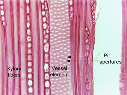
 Fig.
5.1-9. Tangential section of wood of American hornbeam tree (also
called blue beech; Carpinus caroliniana). Cells along the left are xylary
fibers, the cell on the right with the white dots is a vessel element, and its
white dots are the apertures of circular bordered pits. The circular bordered pits of
pine (Fig. 5.1-7) are very large, but these of hornbeam are much more typical of
hardwood trees (that is, dicot trees, trees that are not conifers like pines).
The white spot in the very center of each is the pit aperture, where we are
looking directly into the pit (although the whiteness makes it appear to be a
complete hole, there is a pit membrane that we could see if the microscope
illuminator were turned to be very dim). The light pink halo around each white
spot is the border, where the secondary wall comes up away from the primary
wall. The dark pink lines are the regions where the secondary wall is attached
to the primary wall; in sclereids and libriform fibers, the secondary wall is
attached to the primary wall everywhere, pits have no borders, and all the wall
stains dark pink or red.
Fig.
5.1-9. Tangential section of wood of American hornbeam tree (also
called blue beech; Carpinus caroliniana). Cells along the left are xylary
fibers, the cell on the right with the white dots is a vessel element, and its
white dots are the apertures of circular bordered pits. The circular bordered pits of
pine (Fig. 5.1-7) are very large, but these of hornbeam are much more typical of
hardwood trees (that is, dicot trees, trees that are not conifers like pines).
The white spot in the very center of each is the pit aperture, where we are
looking directly into the pit (although the whiteness makes it appear to be a
complete hole, there is a pit membrane that we could see if the microscope
illuminator were turned to be very dim). The light pink halo around each white
spot is the border, where the secondary wall comes up away from the primary
wall. The dark pink lines are the regions where the secondary wall is attached
to the primary wall; in sclereids and libriform fibers, the secondary wall is
attached to the primary wall everywhere, pits have no borders, and all the wall
stains dark pink or red.