 Fig. 5.1-8.
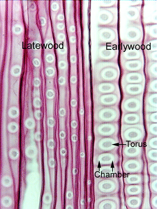
Longitudinal section of wood of pine (Pinus). This section is similar to
that in Fig. 5.1-7 of Zygogynum fibers, but this is a tangential
section of tracheids in pine wood. The large white circles with gray
centers are pits, in this case they are circular bordered
pits. The larger ones
on the right are in the wide earlywood tracheids, the smaller ones on the left
are in the narrow latewood tracheids. In both cases, these pits are vastly
larger than those in the fibers of Zygogynum (Fig. 5.1-7). Pine wood is a
favorite material for demonstrating pits, because these are some of the largest
in existence. Circular bordered pits in tracheids and vessel elements of other
species are narrower, but almost always much larger than the pits of fibers.
Fig. 5.1-8.
Longitudinal section of wood of pine (Pinus). This section is similar to
that in Fig. 5.1-7 of Zygogynum fibers, but this is a tangential
section of tracheids in pine wood. The large white circles with gray
centers are pits, in this case they are circular bordered
pits. The larger ones
on the right are in the wide earlywood tracheids, the smaller ones on the left
are in the narrow latewood tracheids. In both cases, these pits are vastly
larger than those in the fibers of Zygogynum (Fig. 5.1-7). Pine wood is a
favorite material for demonstrating pits, because these are some of the largest
in existence. Circular bordered pits in tracheids and vessel elements of other
species are narrower, but almost always much larger than the pits of fibers.
The gray area in the center of each pine pit is a torus (see page 117 in Chapter 7. Xylem in Plant Anatomy) and the white area is the pit chamber. To see the pit chamber, we are actually looking through some wall material – the border that surrounds the inner aperture of the pit actually overarches the chamber, and we are looking right through it. It seems to be transparent, but that is only because the microscope light is turned bright enough to shine through it. If the illuminator were turned dim enough to see the border, the rest of the slide – which is thicker and denser – would be black.
The vertical lines are the side walls of the tracheids, the walls coming up out of the section toward us. Notice that they do not have pits of any kind.