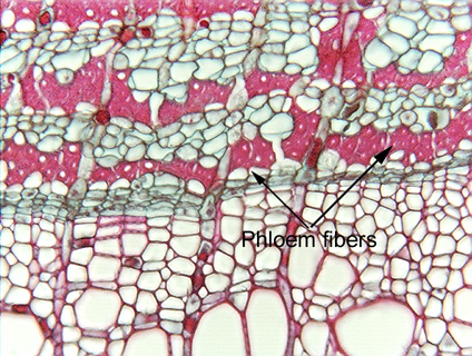
 Fig.
5.3-13. Transverse section of linden tree (Tilia). These two
micrographs show wood (at the bottom) and secondary phloem (the upper part), the
high magnification shows the cambial region where the wood is adjacent to the
phloem. The red bands of cells in the secondary phloem consist of many phloem
fibers, which are so narrow and have such thick walls that only a few
have their lumens visible as small white dots. Secondary phloem is part of the
bark, and bark has several names: “liber” in Latin and “bast” is an
out-of-date term no longer used in English, except when bark fibers are being
described: these phloem fibers can be called “bast
fibers” (used mostly by people interested in textiles). When a
xylary fiber is described as a “libriform fiber,” that means it is as
fiberlike as these bark fibers. Xylary fibers are believed to have evolved from
tracheids, and during the early stages, they still resembled tracheids by having
large pits and thin secondary walls. Those that are as fiber-like as possible --
that is, they resemble the fibers shown here in phloem -- they are said to be
libriform fibers.
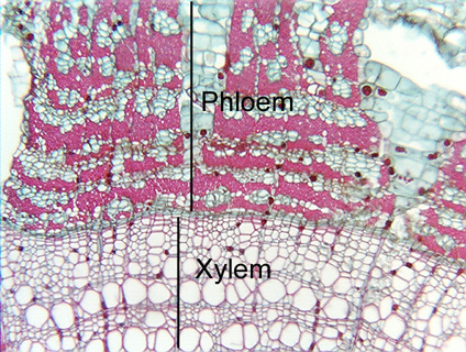
Fig.
5.3-13. Transverse section of linden tree (Tilia). These two
micrographs show wood (at the bottom) and secondary phloem (the upper part), the
high magnification shows the cambial region where the wood is adjacent to the
phloem. The red bands of cells in the secondary phloem consist of many phloem
fibers, which are so narrow and have such thick walls that only a few
have their lumens visible as small white dots. Secondary phloem is part of the
bark, and bark has several names: “liber” in Latin and “bast” is an
out-of-date term no longer used in English, except when bark fibers are being
described: these phloem fibers can be called “bast
fibers” (used mostly by people interested in textiles). When a
xylary fiber is described as a “libriform fiber,” that means it is as
fiberlike as these bark fibers. Xylary fibers are believed to have evolved from
tracheids, and during the early stages, they still resembled tracheids by having
large pits and thin secondary walls. Those that are as fiber-like as possible --
that is, they resemble the fibers shown here in phloem -- they are said to be
libriform fibers.