 Fig. 5.1-2.
Transverse section of wood of Drimys (no common name). The large, more or
less square cells with pink walls are sclerenchyma cells. The white space in the
center of each fiber is the lumen where the protoplast had been located, but
there is no sign of protoplasm now – this sample may have been collected from
a dead piece of lumber or from the dead heartwood of a tree. The narrow white
gaps in the walls are pits, (arrows) regions where the cells did not deposit any
secondary wall interior to the primary wall.
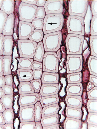
Fig. 5.1-2.
Transverse section of wood of Drimys (no common name). The large, more or
less square cells with pink walls are sclerenchyma cells. The white space in the
center of each fiber is the lumen where the protoplast had been located, but
there is no sign of protoplasm now – this sample may have been collected from
a dead piece of lumber or from the dead heartwood of a tree. The narrow white
gaps in the walls are pits, (arrows) regions where the cells did not deposit any
secondary wall interior to the primary wall.