 Fig.
5.1-6. Transverse section of Aristolochia. This is the region
where two vessel elements contact each
other; the two large white areas are the lumens of the two cells, the space
through which water moves upward. Notice that where the two cell walls touch,
there is a slightly beaded appearance, with faint lens-shaped light areas. The
light areas are the bordered pits. They do not look like the typical
illustrations in any textbook, but that is because sections – even very good,
thin sections – are usually so thick that they contain either the wall behind
the pit, or the wall in front of it, or both, so the pit regions appears to be
just a little lighter in color, a little less stained, rather than a complete
absence of secondary wall. As you can imagine, this can be difficult to see in
most slides made for research. In longitudinal sections, however, the pits are
very obvious.
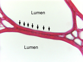
Fig.
5.1-6. Transverse section of Aristolochia. This is the region
where two vessel elements contact each
other; the two large white areas are the lumens of the two cells, the space
through which water moves upward. Notice that where the two cell walls touch,
there is a slightly beaded appearance, with faint lens-shaped light areas. The
light areas are the bordered pits. They do not look like the typical
illustrations in any textbook, but that is because sections – even very good,
thin sections – are usually so thick that they contain either the wall behind
the pit, or the wall in front of it, or both, so the pit regions appears to be
just a little lighter in color, a little less stained, rather than a complete
absence of secondary wall. As you can imagine, this can be difficult to see in
most slides made for research. In longitudinal sections, however, the pits are
very obvious.