 Fig.
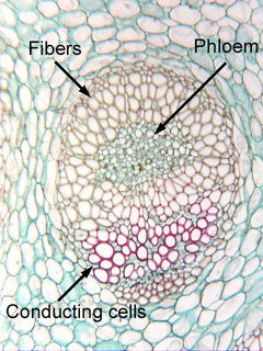
5.1-10. Transverse section through vascular bundle of Cordyline
(no common name). The conducting cells of xylem have thick, red-stained
secondary walls because they are a type of sclerenchyma, so it can be difficult
to distinguish between fibers and conducting cells in transverse sections. In
this vascular bundle, however, the xylem cells have lignified their walls –
and so have become red-stained – whereas the fiber
walls are not lignified and not red-stained. Consequently, it is easy
to tell one type of cell from the other. Notice that the phloem is completely
surrounded by fibers.
Fig.
5.1-10. Transverse section through vascular bundle of Cordyline
(no common name). The conducting cells of xylem have thick, red-stained
secondary walls because they are a type of sclerenchyma, so it can be difficult
to distinguish between fibers and conducting cells in transverse sections. In
this vascular bundle, however, the xylem cells have lignified their walls –
and so have become red-stained – whereas the fiber
walls are not lignified and not red-stained. Consequently, it is easy
to tell one type of cell from the other. Notice that the phloem is completely
surrounded by fibers.