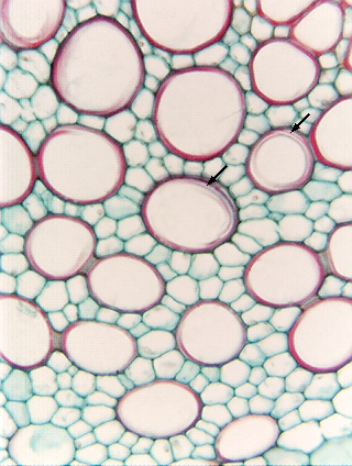
 Fig.
11.5-9. Transverse section of parsnip stem (Pastinaca
sativa). The xylem parenchyma is abundant and almost completely surrounds
every vessel – there are few contact faces between vessels. Instead,
the great majority of the surface area of each vessel element faces a
xylem parenchyma cell. The vessel elements here have annular and
helical secondary walls (arrows), which means that most of the surface area of
each vessel is just primary wall, directly abutting the primary walls of
parenchyma cells.
Fig.
11.5-9. Transverse section of parsnip stem (Pastinaca
sativa). The xylem parenchyma is abundant and almost completely surrounds
every vessel – there are few contact faces between vessels. Instead,
the great majority of the surface area of each vessel element faces a
xylem parenchyma cell. The vessel elements here have annular and
helical secondary walls (arrows), which means that most of the surface area of
each vessel is just primary wall, directly abutting the primary walls of
parenchyma cells.