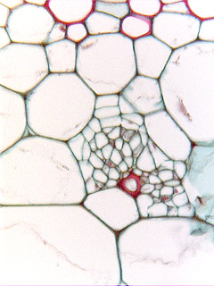
 Fig.
11.5-12. Transverse section of tobacco stem (Nicotiana
tabacum). This is the internal phloem of a bicollateral vascular bundle:
protoxylem is at the top of the micrograph, and the ordinary phloem would be
high above that, far out of view. The phloem here – internal phloem – is
located in the pith, and its sieve tube members and companion cells are easy to
identify. An unusual feature is that it has a fiber. Note
that the internal phloem is very close to the protoxylem, only one parenchyma
cell away. Internal phloem is always close to the xylem, it is not
located deep in the pith.
Fig.
11.5-12. Transverse section of tobacco stem (Nicotiana
tabacum). This is the internal phloem of a bicollateral vascular bundle:
protoxylem is at the top of the micrograph, and the ordinary phloem would be
high above that, far out of view. The phloem here – internal phloem – is
located in the pith, and its sieve tube members and companion cells are easy to
identify. An unusual feature is that it has a fiber. Note
that the internal phloem is very close to the protoxylem, only one parenchyma
cell away. Internal phloem is always close to the xylem, it is not
located deep in the pith.