 Fig.
11.3-4.
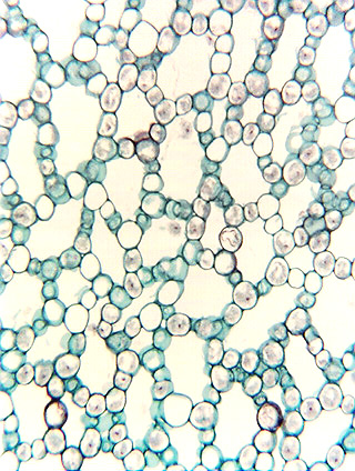
Transverse section of stem cortex in sweetflag (Acorus). The cortex of
sweetflag becomes several millimeters thick, and this area is much closer to the
center of the stem than that of the previous micrograph. The intercellular
spaces here are even larger, and it is obvious that the “walls” of each
space consist of just a single layer of cortex cells: each cell is exposed to an
intercellular space on several sides. These spaces are probably schizogenous –
the young, developing tissue would have been compact but then at some point
during enlargement, the cells pulled apart from each other. We
know very little about the underlying mechanism – were the middle lamellas
torn apart in certain areas or were they digested enzymatically by the cells?
What mechanism determined which areas of middle lamella would be ruptured and
which would remain intact?
Fig.
11.3-4.
Transverse section of stem cortex in sweetflag (Acorus). The cortex of
sweetflag becomes several millimeters thick, and this area is much closer to the
center of the stem than that of the previous micrograph. The intercellular
spaces here are even larger, and it is obvious that the “walls” of each
space consist of just a single layer of cortex cells: each cell is exposed to an
intercellular space on several sides. These spaces are probably schizogenous –
the young, developing tissue would have been compact but then at some point
during enlargement, the cells pulled apart from each other. We
know very little about the underlying mechanism – were the middle lamellas
torn apart in certain areas or were they digested enzymatically by the cells?
What mechanism determined which areas of middle lamella would be ruptured and
which would remain intact?