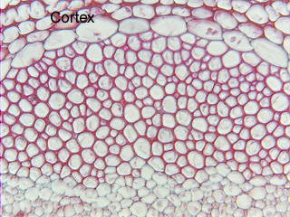
 Fig.
11.5-15. Phloem of ragweed (Ambrosia). This
micrograph shows the primary phloem fiber cap. This is a relatively
large cap, containing many fibers. They are often smaller in other species, even
consisting of as few as one or two fibers in any particular transverse section.
Although this is a low magnification, you may be able to see a few nuclei and
other cell contents – at least at this stage, these fibers have remained alive
although they have completed the deposition and lignification of their secondary
wall.
Fig.
11.5-15. Phloem of ragweed (Ambrosia). This
micrograph shows the primary phloem fiber cap. This is a relatively
large cap, containing many fibers. They are often smaller in other species, even
consisting of as few as one or two fibers in any particular transverse section.
Although this is a low magnification, you may be able to see a few nuclei and
other cell contents – at least at this stage, these fibers have remained alive
although they have completed the deposition and lignification of their secondary
wall.