 Fig.
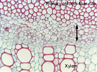
11.5-14. Transverse section of ragweed stem (Ambrosia). This
micrograph shows the phloem of a dicot vascular bundle. The region indicated by
the double-headed arrow contains the conducting cells – the sieve tube members
– but the phloem consists of both that and the fiber cap as well. This is rather a large amount of phloem,
but still the entire band of sieve tube members and companion cells is no wider
than one of the larger vessels.
Fig.
11.5-14. Transverse section of ragweed stem (Ambrosia). This
micrograph shows the phloem of a dicot vascular bundle. The region indicated by
the double-headed arrow contains the conducting cells – the sieve tube members
– but the phloem consists of both that and the fiber cap as well. This is rather a large amount of phloem,
but still the entire band of sieve tube members and companion cells is no wider
than one of the larger vessels.