Up
Dicot stem
Monocot stem
Broad pith
Weak stem
Monocot fiber sheaths
Ordinary cortex
Aerenchyma hypodermis
Aerenchyma cortex
Aerenchyma cortex 2
Stem endodermis
Palisade cortex
Cortical bundle
Capped cortical bundles
Collapsible cortex
Perimedullary fibers
Conjunctive tissue, paren.
Torn pith
Hollow pith
Medullary bundles
Typical dicot bundle
Vascular ring
Typical monocot bundle
Amphivasal bundle
Corn vascular bundle
Clintonia bundles
Protoxylem
Metaxylem
Metaxylem parenchyma
Metaxylem fibers
Internal phloem
Internal phloem, mag
Developing metaxylem
Primary phloem
Phloem fiber cap
Developing fibers
| |
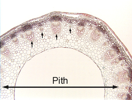 Fig.
11.4-4. Transverse section of delphinium stem (Delphinium ajacis).
This delphinium stem is a good example of a stem in which the pith breaks down.
The pith extends from the innermost protoxylem on one side of the stem to the
innermost protoxylem on the other side, as far as the arrow extends. Not
all the pith has been torn apart; the perimedullary region – a layer about
five or six cells thick -- has elongated enough to keep up with the expansion of
the rest of the stem tissues, so it is intact. Fig.
11.4-4. Transverse section of delphinium stem (Delphinium ajacis).
This delphinium stem is a good example of a stem in which the pith breaks down.
The pith extends from the innermost protoxylem on one side of the stem to the
innermost protoxylem on the other side, as far as the arrow extends. Not
all the pith has been torn apart; the perimedullary region – a layer about
five or six cells thick -- has elongated enough to keep up with the expansion of
the rest of the stem tissues, so it is intact.
The small arrows indicate four of the many vascular bundles. Some bundles
are so large that the vessels in their xylem are visible even at this low
magnification, other bundles are so small the entire bundle is scarcely visible.
Many monocots also have hollow stems (grasses for example), and often so much
conjunctive tissue breaks down that there is only enough room left for one ring
of bundles. In such monocot stems, the atactostele can then be easily mistaken
for the eustele of a dicot. If your anatomy lab uses wheat (Triticum) as
teaching slides, you may run into this (hint to students – professors like to
use this on lab tests).
|