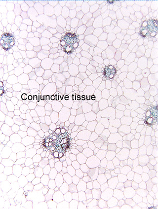
 Fig.
11.4-2. Transverse section of corn stem (Zea
mays). The vascular bundles of monocots stems typically occurs in a complex
three dimensional pattern embedded in a matrix that is called conjunctive
tissue. Conjunctive tissue may be either parenchyma as in
this corn, or it can consist of fibers whose walls can become extremely thick.
As you might imagine, parenchymatous conjunctive tissue occurs in monocot stems
that are soft – such as corn, canna lilies, gingers, and bulbs like onions –
whereas a fibrous conjunctive tissue occurs in larger, harder monocot stems like
bamboo, agaves, and many species of palm trees.
Fig.
11.4-2. Transverse section of corn stem (Zea
mays). The vascular bundles of monocots stems typically occurs in a complex
three dimensional pattern embedded in a matrix that is called conjunctive
tissue. Conjunctive tissue may be either parenchyma as in
this corn, or it can consist of fibers whose walls can become extremely thick.
As you might imagine, parenchymatous conjunctive tissue occurs in monocot stems
that are soft – such as corn, canna lilies, gingers, and bulbs like onions –
whereas a fibrous conjunctive tissue occurs in larger, harder monocot stems like
bamboo, agaves, and many species of palm trees.
In this micrograph, we can see eight vascular bundles, but if we could somehow make the conjunctive tissue perfectly transparent so that we could see far down into this stem, almost certainly we would see that some of them come together and join to be a singe bundle, others might branch into two and so on, such that they form an interconnected network, not just a set of bundles each isolated from the others.