 Fig.
10.3-7. Magnification of fig leaf (Ficus).
This micrograph shows two stomata. The one on the right has been cut
parallel to the stomatal pore, and we see one of the guard cells in longitudinal
section. The guard cell appears to be a cutin-covered bridge that
blocks the substomatal chamber from the air outside the leaf, but actually the
stomatal pore is present just out of the plane of sectioning.
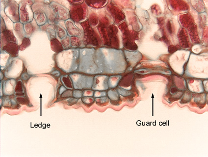
Fig.
10.3-7. Magnification of fig leaf (Ficus).
This micrograph shows two stomata. The one on the right has been cut
parallel to the stomatal pore, and we see one of the guard cells in longitudinal
section. The guard cell appears to be a cutin-covered bridge that
blocks the substomatal chamber from the air outside the leaf, but actually the
stomatal pore is present just out of the plane of sectioning.
The stoma on the left is also cut parallel to the guard cell, but the section is almost at the pore – it has just grazed a guard cell ledge, so the stoma appears to be blocked. Notice how small the two guard cells appear to be: those are actually the two ends of the same cell – the cell is crescent shaped and the section has just barely caught the two ends. If the section were a little deeper into the guard cell, it would look like the one on the right.