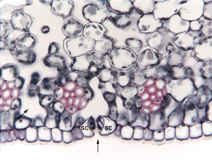
 Fig.
10.3-4. Transverse section of leaf Dracaena
fragrans (no common name). This is a very typical
view of stomata, visible in a transverse section of a leaf. The
vertical arrow indicates a stomatal complex: there are two subsidiary cells (sc)
visible here, and two guard cells that look like little more than smudges on the
inner sides of the subsidiary cells. Above the stoma is a white space with no
cells, that is a substomatal cavity that is open enough that the carbon dioxide
molecules will diffuse quickly away from the stomatal pore and be less likely to
accidentally diffuse back out of the stoma. The diagonal arrow indicates another
open area; undoubtedly it too has a stoma that opens into it, but that stoma
must be a little farther back in the epidermis or it was a little farther
forward and has been cut away in a preceding section. And almost certainly, that
substomatal chamber is connected to the other chamber whose stoma is indicated
by the arrow: the chambers are part of an interconnected space.
Fig.
10.3-4. Transverse section of leaf Dracaena
fragrans (no common name). This is a very typical
view of stomata, visible in a transverse section of a leaf. The
vertical arrow indicates a stomatal complex: there are two subsidiary cells (sc)
visible here, and two guard cells that look like little more than smudges on the
inner sides of the subsidiary cells. Above the stoma is a white space with no
cells, that is a substomatal cavity that is open enough that the carbon dioxide
molecules will diffuse quickly away from the stomatal pore and be less likely to
accidentally diffuse back out of the stoma. The diagonal arrow indicates another
open area; undoubtedly it too has a stoma that opens into it, but that stoma
must be a little farther back in the epidermis or it was a little farther
forward and has been cut away in a preceding section. And almost certainly, that
substomatal chamber is connected to the other chamber whose stoma is indicated
by the arrow: the chambers are part of an interconnected space.