 Fig.
10.3-6.
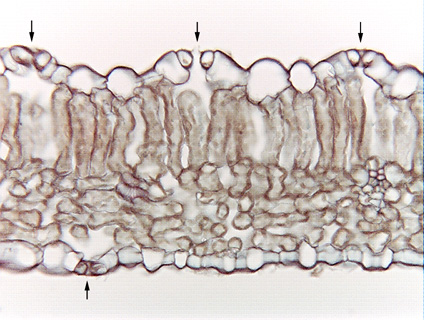
Transverse section of parsnip leaf (Pastinaca sativa). Four stomata are
visible here: the center one in the upper epidermis is cut through its center,
showing the two guard cells and two subsidiary cells and the stomatal pore. The
stoma on the right seems to have some material across the top of the stomatal
pore: actually, the
section has caught a little of the end of the stomatal pore, where
the two guard cells are in contact even when the pore is open. The pore only
appears to be blocked. The stoma on the left in the upper epidermis, and the one
in the lower epidermis have been cut even closer to the end of the guard cells
-- none of the stomatal pore is visible in either of them. We can recognize that
they are stomata because the guard cells are smaller than ordinary epidermis
cells and each has a substomatal chamber interior to it. The
upper left stoma looks strange as well because it is oriented at an angle to the
section: usually, we only photograph those stomata that have -- by
luck -- been cut in perfect transverse section and right through the center.
When you are examining slides in lab, you will see them cut at every possible
orientation.
Fig.
10.3-6.
Transverse section of parsnip leaf (Pastinaca sativa). Four stomata are
visible here: the center one in the upper epidermis is cut through its center,
showing the two guard cells and two subsidiary cells and the stomatal pore. The
stoma on the right seems to have some material across the top of the stomatal
pore: actually, the
section has caught a little of the end of the stomatal pore, where
the two guard cells are in contact even when the pore is open. The pore only
appears to be blocked. The stoma on the left in the upper epidermis, and the one
in the lower epidermis have been cut even closer to the end of the guard cells
-- none of the stomatal pore is visible in either of them. We can recognize that
they are stomata because the guard cells are smaller than ordinary epidermis
cells and each has a substomatal chamber interior to it. The
upper left stoma looks strange as well because it is oriented at an angle to the
section: usually, we only photograph those stomata that have -- by
luck -- been cut in perfect transverse section and right through the center.
When you are examining slides in lab, you will see them cut at every possible
orientation.