 Fig.
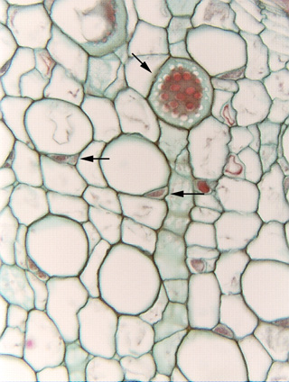
8.2-7. High magnification of cucumber phloem. In the upper part of
the micrograph is a sieve plate (diagonal
arrow) in which the sieve pores (white areas) are very large and easily visible.
This is typical of cucurbits but in most other plants they are much small and
much more difficult to see. The red discoloration is the P-protein
plug, which is not completely occluding all the pores of the plate.
Fig.
8.2-7. High magnification of cucumber phloem. In the upper part of
the micrograph is a sieve plate (diagonal
arrow) in which the sieve pores (white areas) are very large and easily visible.
This is typical of cucurbits but in most other plants they are much small and
much more difficult to see. The red discoloration is the P-protein
plug, which is not completely occluding all the pores of the plate.
The two horizontal arrows point out especially nice pairs of sieve tube members and companion cells. Notice that in both cases, the companion cell has the appearance of being just a corner of the sieve tube member: if the wall between the sieve tube member and its associated companion cell were removed, the resulting cell would have a very natural size and shape.