 Fig.
8.1-4b. Transverse section of parsnip (Pastinaca)
stem. The sieve tube members (several marked with s) in this dicot phloem are
relatively easy to recognize because they are rather large and have the typical
empty appearance. However, notice that many of the cells in the cortex have a
similar size and appearance. In the phloem, there are many small, cytoplasmic
cells that appear to be companion cells, and these are completely absent from
the cortex. In a few of the cases here, it seems possible to make a guess as to
which companion cell might be associated with which sieve tube member, but it
would only be a guess -- it is necessary to use transmission electron microscopy
to examine the primary pit field/sieve area connections to be certain.
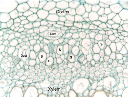
Fig.
8.1-4b. Transverse section of parsnip (Pastinaca)
stem. The sieve tube members (several marked with s) in this dicot phloem are
relatively easy to recognize because they are rather large and have the typical
empty appearance. However, notice that many of the cells in the cortex have a
similar size and appearance. In the phloem, there are many small, cytoplasmic
cells that appear to be companion cells, and these are completely absent from
the cortex. In a few of the cases here, it seems possible to make a guess as to
which companion cell might be associated with which sieve tube member, but it
would only be a guess -- it is necessary to use transmission electron microscopy
to examine the primary pit field/sieve area connections to be certain.
This phloem also has two small secretory ducts.