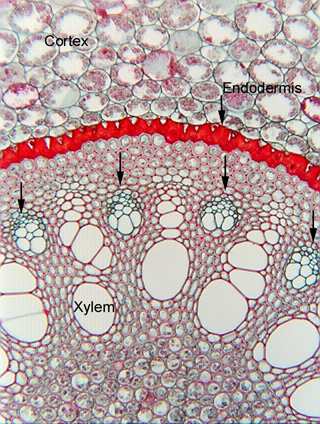
 Fig.
8.1-7a. Transverse section of root of catclaw (Smilax).
In the roots of many monocots, the cells that make up the matrix (called
conjunctive tissue) between the tracheary elements and the sieve tube members
are thick-walled fibers. In comparison to their darkly stained, red walls, the
thin-walled, green sieve tube members and companion cells (arrows) are easy to
identify. Even at this low magnification, we know immediately where all the
phloem is. Notice that it
alternates with the masses of tracheary elements, near the outer edge of the
xylem, and just a little interior to the endodermis. Learning the
location here where it is easy to see will help you find the phloem in those
monocot roots that have a parenchymatous conjunctive tissue.
Fig.
8.1-7a. Transverse section of root of catclaw (Smilax).
In the roots of many monocots, the cells that make up the matrix (called
conjunctive tissue) between the tracheary elements and the sieve tube members
are thick-walled fibers. In comparison to their darkly stained, red walls, the
thin-walled, green sieve tube members and companion cells (arrows) are easy to
identify. Even at this low magnification, we know immediately where all the
phloem is. Notice that it
alternates with the masses of tracheary elements, near the outer edge of the
xylem, and just a little interior to the endodermis. Learning the
location here where it is easy to see will help you find the phloem in those
monocot roots that have a parenchymatous conjunctive tissue.
See Fig. 8.1-7b for a higher magnification view.