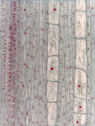
 Fig.
6.8-3. Longitudinal section through the zone of elongation just
proximal to the root apex of cattail (Typha). Notice the tremendous
differences in cells size. The very large cells in the two columns in the center
will become vessel elements in the xylem, the surrounding smaller cells will
differentiate into a variety of vascular tissues. The main point illustrated
here is that as a given block of living tissue moves away from the apical
meristem and expands, it will form a few large cells if cell division is slow or
many small cells if it is rapid. For example, two small cubes of meristem tissue
measuring 10mm
on each side might both expand to become blocks of mature tissue measuring 1000mm
on each side, but one may be divided up into many small cells, the other into
just a few large cells. It is easy to assume that a large cell is larger because
it is growing faster than adjacent small cells, but that would be incorrect.
Fig.
6.8-3. Longitudinal section through the zone of elongation just
proximal to the root apex of cattail (Typha). Notice the tremendous
differences in cells size. The very large cells in the two columns in the center
will become vessel elements in the xylem, the surrounding smaller cells will
differentiate into a variety of vascular tissues. The main point illustrated
here is that as a given block of living tissue moves away from the apical
meristem and expands, it will form a few large cells if cell division is slow or
many small cells if it is rapid. For example, two small cubes of meristem tissue
measuring 10mm
on each side might both expand to become blocks of mature tissue measuring 1000mm
on each side, but one may be divided up into many small cells, the other into
just a few large cells. It is easy to assume that a large cell is larger because
it is growing faster than adjacent small cells, but that would be incorrect.