 Fig.
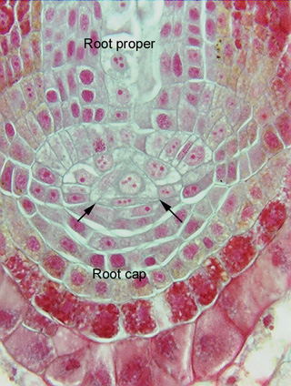
6.8-1b. Magnification of the root apical meristem of a fern. The single
apical cell is very prominent in this micrograph, or at least its
nucleus is. Notice the thin, flat cell between the two arrows: it is a progeny
cell of the apical cell, but it was cut off to the forward side of the apical
cell. This flat cell will continue to divide, and its progeny cells will become
root cap cells. The cells cut off from the other three faces of the apical cell
(only two of those three faces are visible here) will all contribute to the root
proper. You can even see that these progeny cells divide so as to produce sets
of brick-shaped cells that lie parallel to each other.
Fig.
6.8-1b. Magnification of the root apical meristem of a fern. The single
apical cell is very prominent in this micrograph, or at least its
nucleus is. Notice the thin, flat cell between the two arrows: it is a progeny
cell of the apical cell, but it was cut off to the forward side of the apical
cell. This flat cell will continue to divide, and its progeny cells will become
root cap cells. The cells cut off from the other three faces of the apical cell
(only two of those three faces are visible here) will all contribute to the root
proper. You can even see that these progeny cells divide so as to produce sets
of brick-shaped cells that lie parallel to each other.
The abundant red-stained material is tannin, which is extremely common in ferns.