Up
Typical collen.
Developing collen.
Mature collen.
Thickened corners
Column in cortex
Nuclei
Lamellar, sparse
Lamellar, abundant
Pumpkin petiole
Birch petiole
Leaf vein
Leaf hypodermis
| |
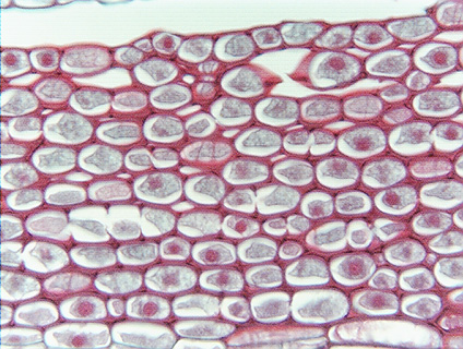 Fig. 4.1-8.
Transverse section of stem of elder (Sambucus). This is an unusual sample
of collenchyma in that there is a large amount of lamellar
collenchyma and it has a large number of intercellular spaces. This
particular mass of collenchyma was located below a lenticel, a patch of loose
cork cells that permits oxygen to diffuse into the living tissues below the
bark. The intercellular spaces in this collenchyma permit oxygen to penetrate to
the tissues below. These collenchyma cells have prominent protoplasts (gray;
plasmolyzed during processing), and there are so many nuclei (red) present that
these must be short cells. Fig. 4.1-8.
Transverse section of stem of elder (Sambucus). This is an unusual sample
of collenchyma in that there is a large amount of lamellar
collenchyma and it has a large number of intercellular spaces. This
particular mass of collenchyma was located below a lenticel, a patch of loose
cork cells that permits oxygen to diffuse into the living tissues below the
bark. The intercellular spaces in this collenchyma permit oxygen to penetrate to
the tissues below. These collenchyma cells have prominent protoplasts (gray;
plasmolyzed during processing), and there are so many nuclei (red) present that
these must be short cells.
It is unusual that cell walls are stained red rather than green. If this
material was stained with Safranin and Fast Green, then the red color would
indicate that these collenchyma cells have lignified their walls. That is fairly
common, especially in collenchyma that occurs in long-lived organs like a stem.
On the other hand, it may be that this slide was stained with some other, less
common stain that gives an unlignified wall a red color rather than a green
color.
|