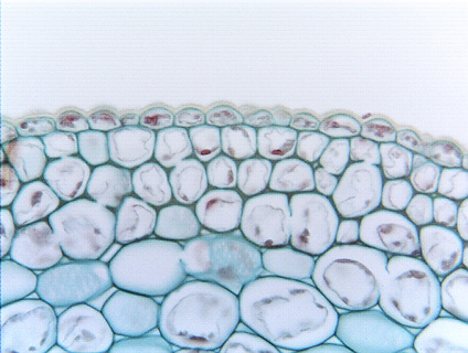
 Fig. 4.1-2.
Transverse section of stem of ivy (Hedera helix). This micrograph shows
three layers of collenchyma cells in an early stage of development, before the
corners have become very thick. It is possible to see that the
cells actually are angular, not round, and with no intercellular
spaces between the collenchyma cells themselves. As development continues, the
corners would thicken even more and cell lumens would become round as in Fig.
4.1-1. The uppermost layer is the epidermis, and the lower, larger cells are
ordinary cortex cells. Notice that there are intercellular spaces between the
innermost collenchyma cells and their neighboring ordinary cortex parenchyma
cells. Also, the innermost collenchyma cells have thickened corners where they
abut other collenchyma cells but not where they abut parenchyma cells.
Fig. 4.1-2.
Transverse section of stem of ivy (Hedera helix). This micrograph shows
three layers of collenchyma cells in an early stage of development, before the
corners have become very thick. It is possible to see that the
cells actually are angular, not round, and with no intercellular
spaces between the collenchyma cells themselves. As development continues, the
corners would thicken even more and cell lumens would become round as in Fig.
4.1-1. The uppermost layer is the epidermis, and the lower, larger cells are
ordinary cortex cells. Notice that there are intercellular spaces between the
innermost collenchyma cells and their neighboring ordinary cortex parenchyma
cells. Also, the innermost collenchyma cells have thickened corners where they
abut other collenchyma cells but not where they abut parenchyma cells.