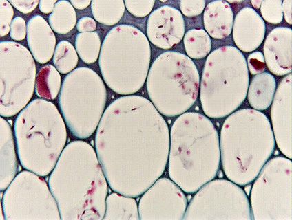
 Fig.
4.1-3. Transverse section of petiole of water lily (Nymphaea).
This is the region where fully-formed collenchyma
(at the top of the micrograph) grades into ordinary parenchyma (at the bottom).
Near the top, it is possible to see the middle lamella between several of the
cells, and because the middle lamella is straight, so is the outer surface of
the cell wall – these cells really are angular, not spherical. Notice that
there are intercellular spaces between the parenchyma cells and collenchyma
cells and even between some of the innermost collenchyma cells.
Fig.
4.1-3. Transverse section of petiole of water lily (Nymphaea).
This is the region where fully-formed collenchyma
(at the top of the micrograph) grades into ordinary parenchyma (at the bottom).
Near the top, it is possible to see the middle lamella between several of the
cells, and because the middle lamella is straight, so is the outer surface of
the cell wall – these cells really are angular, not spherical. Notice that
there are intercellular spaces between the parenchyma cells and collenchyma
cells and even between some of the innermost collenchyma cells.