Up
Primary xylem
Oak wood
Leaf vein
Vein ends
Bean seed
Pine tracheids, xs
Fern TE, xs
Fern, TE, mag
Annular walls
Annular, stretched
Annular, narrow
Scalariform walls
Scalar., narrow
CBP, pine
CBP, dicot
CBP, irregular
Contact faces
Pits, side view
CBP, pine, xs
CBP,angio, xs
CBP, fern, xs
Contact face, xs
Simple perf. plate 1
Simple perf. plate 2
Pitted perf. plate
Perf. plate & helix
Perf. plate, face
Perf. plate, mag
Perf. plate, section
Perf. plate rim
Perf. plate & wall
Scalariform Per plate
Primary xylem
Vessel sizes
Fern TE
Pine needle
VE precursor, ls
Protoxylem
9 Contact faces
VE precursor, xs
Precursor 2
Torn vessel
Torn vessel 2
| |
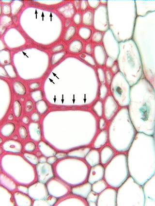 Fig.
7.2-13. Transverse section of cosmos (Cosmos) stem. These
vessel elements are interconnected by circular
bordered pits, visible here in transverse section. Compare this view
to the large circular bordered pits in pine (Fig. 7.2-11) or to the scalariform
pits of grape (Fig. 7.2-12a). We can tell that these must be circular bordered
pits because they do not extend all the way across the contact face: the arrows
indicate the individual pit chambers, and between each arrow are the borders of
the pits. Fig.
7.2-13. Transverse section of cosmos (Cosmos) stem. These
vessel elements are interconnected by circular
bordered pits, visible here in transverse section. Compare this view
to the large circular bordered pits in pine (Fig. 7.2-11) or to the scalariform
pits of grape (Fig. 7.2-12a). We can tell that these must be circular bordered
pits because they do not extend all the way across the contact face: the arrows
indicate the individual pit chambers, and between each arrow are the borders of
the pits.
This is not really an easy view for beginners to see. You have to examine
your slides carefully at high power (a 25x or 40x objective), and the sections
must have been cut well to give a clean surface. However, with just a little
practice, it is not too difficult to see this beaded appearance. You need to
worry about this only if you are examining a tissue transverse section and want
to determine if a cell is a fiber of a tracheary element: if there is any beaded
appearance to the wall at all, it must contain bordered pits. Fibers will have
very narrow pits that are either definitely much more slender than these or so
slender you almost cannot see them – if the wall appears completely uniform,
it probably is a fiber.
|