 Fig.
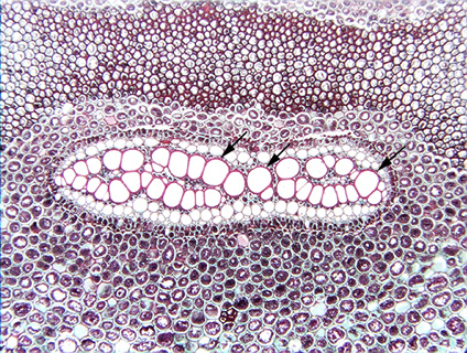
7.1-7a. Transverse section of rhizome of a fern (Pteridium).
The center of the section is occupied by a plate of large cells with thick,
red-stained walls. What are they? They are shown in higher magnification in
7.1-7b, but that will not help. A few years ago, we would have confidently
stated that because this is a fern, these are almost certainly tracheids. Pteridium
was one of just four ferns known to have vessels. But in the last few years
(since the late 1990s), Dr. S. Carlquist and E. Schneider have been discovering
that in a
considerable number of ferns, structures that were thought to be pits are
actually perforations -- the pit membrane is digested away during
cell maturation. Only by using scanning electron microscopy (SEM) is it possible
to see that the pit membrane is missing. For an introduction to their research,
see Schneider, D. L., and S. Carlquist. 2000. SEM studies on vessels in ferns.
American Journal of Botany 87: 176-181.
Fig.
7.1-7a. Transverse section of rhizome of a fern (Pteridium).
The center of the section is occupied by a plate of large cells with thick,
red-stained walls. What are they? They are shown in higher magnification in
7.1-7b, but that will not help. A few years ago, we would have confidently
stated that because this is a fern, these are almost certainly tracheids. Pteridium
was one of just four ferns known to have vessels. But in the last few years
(since the late 1990s), Dr. S. Carlquist and E. Schneider have been discovering
that in a
considerable number of ferns, structures that were thought to be pits are
actually perforations -- the pit membrane is digested away during
cell maturation. Only by using scanning electron microscopy (SEM) is it possible
to see that the pit membrane is missing. For an introduction to their research,
see Schneider, D. L., and S. Carlquist. 2000. SEM studies on vessels in ferns.
American Journal of Botany 87: 176-181.