 Fig.
7.2-10. Longitudinal section through a vessel element of corn. This
is a view you might see in your own laboratory material, but only if you focus
at the very top of the section at high power. Histological slides, purchased
from companies like Triarch or Carolina Biological, or made in your own lab, are
usually between 8mm
and 15mm.
That is thick compared to the plane of focus of the microscope (the level within
the specimen where the microscope is obtaining the most precisely focused
image). As you adjust the fine focus knob, you are moving the stage to bring
different levels of the specimen into the plane of focus. To get an image like
the one here, you must lower the specimen so much that the very top cut surface
of the section – right where the microtome knife passed – is in the plane of
focus.
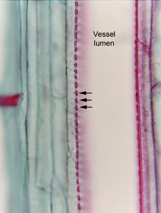
Fig.
7.2-10. Longitudinal section through a vessel element of corn. This
is a view you might see in your own laboratory material, but only if you focus
at the very top of the section at high power. Histological slides, purchased
from companies like Triarch or Carolina Biological, or made in your own lab, are
usually between 8mm
and 15mm.
That is thick compared to the plane of focus of the microscope (the level within
the specimen where the microscope is obtaining the most precisely focused
image). As you adjust the fine focus knob, you are moving the stage to bring
different levels of the specimen into the plane of focus. To get an image like
the one here, you must lower the specimen so much that the very top cut surface
of the section – right where the microtome knife passed – is in the plane of
focus.
What we are seeing here (at the arrows) is the side wall of a vessel element coming right up out of the section at us. The small red “beads” are the portions of secondary wall. If this were a vessel element with annular secondary walls, these would be the cut ends of the rings. Actually, this vessel element has circular bordered pits (I know that because I focused down to see the back wall before I took the micrograph), so each of the spaces between the red beads is a pit chamber. Note that each bead is somewhat triangle-shaped with the apex of the triangle point toward the primary wall; the two sides that face the pit chamber are the borders of the pit.
The wall on the right side of the cell has the same structure, but it is a tiny bit lower than the left side, so it is too far out of the plane of focus to see it. If we were to refocus on the right side, the left side would be out of focus.