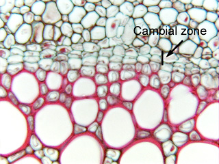
 Fig. 3.2-5. Transverse
section of stem of angelica (Angelica, an ornamental herb in the parsley
family) The large red-stained cells at the bottom of the micrograph are xylem
cells, the larger ones at the top are phloem cells. The band of flat, cuboidal
or box-like cells in between xylem and phloem are vascular
cambium cells, another type of synthetic parenchyma. Although these
appear to be small cells, like those of the shoot apical meristem in Fig. 3.2-4,
the fact that so few nuclei are visible indicates something must be not quite as
it appears. These are actually extremely long cells that have been cut in
transverse section – most cells are probably about 100 to 200mm
long and this section is only about 10mm
thick, so there is a 10 in 100 to 200 (1 in 10 to 20) chance of cutting through
one of these cells in an area where a nucleus happens to be.
Fig. 3.2-5. Transverse
section of stem of angelica (Angelica, an ornamental herb in the parsley
family) The large red-stained cells at the bottom of the micrograph are xylem
cells, the larger ones at the top are phloem cells. The band of flat, cuboidal
or box-like cells in between xylem and phloem are vascular
cambium cells, another type of synthetic parenchyma. Although these
appear to be small cells, like those of the shoot apical meristem in Fig. 3.2-4,
the fact that so few nuclei are visible indicates something must be not quite as
it appears. These are actually extremely long cells that have been cut in
transverse section – most cells are probably about 100 to 200mm
long and this section is only about 10mm
thick, so there is a 10 in 100 to 200 (1 in 10 to 20) chance of cutting through
one of these cells in an area where a nucleus happens to be.