 Fig.
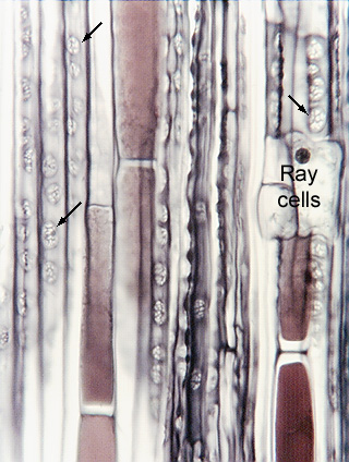
16.1-3a and b. Radial section of pine bark. The large micrograph
shows the simple nature of secondary phloem in conifers.
The tall cells with dark contents are tannin cells, all the other tall cells are
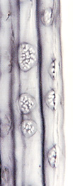
sieve cells (the arrows indicate a few sieve areas, which are shown in higher
magnification in the small micrograph). Each white dot in the sieve areas is an
individual sieve pore. Pine wood is similarly simple, having just tracheids and
a few resin canals.
Fig.
16.1-3a and b. Radial section of pine bark. The large micrograph
shows the simple nature of secondary phloem in conifers.
The tall cells with dark contents are tannin cells, all the other tall cells are
sieve cells (the arrows indicate a few sieve areas, which are shown in higher
magnification in the small micrograph). Each white dot in the sieve areas is an
individual sieve pore. Pine wood is similarly simple, having just tracheids and
a few resin canals.
The microtome knife caught the very
edge of a ray, so three ray cells (and a nucleus) are visible as well.