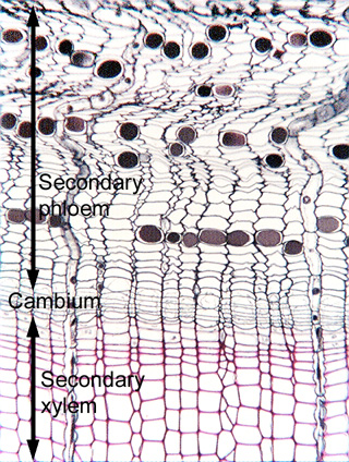
 Fig.
16.1-1. Transverse section of pine (Pinus).
This is a low magnification view for orientation. Wood (secondary xylem) is at
the bottom of the micrograph (the inner part of the tree trunk), the cambial
zone runs across the center, and the secondary phloem is all the tissue above
the cambium (to the outside of the cambium). The fusiform initials that produce
rows of tracheids in the secondary xylem also produce sieve cells in the
secondary phloem (the cambium is bidirectional), and likewise, ray initials
produce rays in phloem as well as xylem. The cells with darkly stained contents
in the phloem are tannin cells. Although
sieve cells are abundant here, they are virtually impossible to identify in
transverse section – the sieve areas cannot be seen in this
micrograph. Albuminous cells occur in the rays; they are not sister cells to the
sieve cells and are not even produced by the fusiform initials (see pages 343
and 344 in Plant Anatomy (Mauseth)).
Fig.
16.1-1. Transverse section of pine (Pinus).
This is a low magnification view for orientation. Wood (secondary xylem) is at
the bottom of the micrograph (the inner part of the tree trunk), the cambial
zone runs across the center, and the secondary phloem is all the tissue above
the cambium (to the outside of the cambium). The fusiform initials that produce
rows of tracheids in the secondary xylem also produce sieve cells in the
secondary phloem (the cambium is bidirectional), and likewise, ray initials
produce rays in phloem as well as xylem. The cells with darkly stained contents
in the phloem are tannin cells. Although
sieve cells are abundant here, they are virtually impossible to identify in
transverse section – the sieve areas cannot be seen in this
micrograph. Albuminous cells occur in the rays; they are not sister cells to the
sieve cells and are not even produced by the fusiform initials (see pages 343
and 344 in Plant Anatomy (Mauseth)).
Much of the outer part of the phloem rows are undulate: as the sieve cells stop functioning and collapse, axial tissue contracts into this irregular pattern. Because most ray cells do not collapse, rays retain their size and consequently are forced into folds by the collapsing sieve cells.