 Fig.
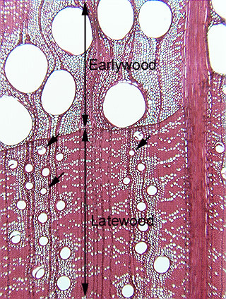
15.3-9a and b. Transverse section of oak wood (Quercus). Oak
wood is a complex dicot wood because its
axial system has fibers, parenchyma, and several sizes of vessels. As
is typical, the micrograph is oriented as if the vascular cambium were above the
top of the computer screen, so the earlywood in the upper part of the picture is
part of the annual ring that was produced the year after the latewood of the
lower part. The earlywood vessels have a diameter six or seven times greater
than the diameter of the narrow vessels of the latewood, and remember that a
vessel’s conducting capacity is proportional to its radius taken to the fourth
power (r4)(see page 113 in Plant Anatomy (Mauseth)): the
conducting capacity of the large vessels is hundreds of times greater than that
of the narrow ones, not just six or seven times greater.
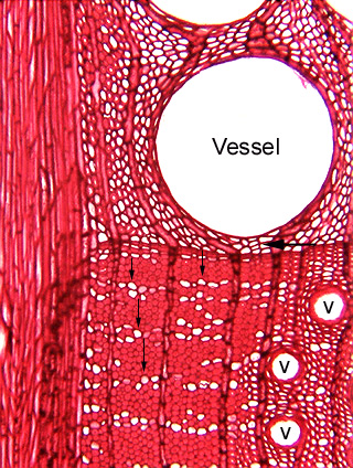
Fig.
15.3-9a and b. Transverse section of oak wood (Quercus). Oak
wood is a complex dicot wood because its
axial system has fibers, parenchyma, and several sizes of vessels. As
is typical, the micrograph is oriented as if the vascular cambium were above the
top of the computer screen, so the earlywood in the upper part of the picture is
part of the annual ring that was produced the year after the latewood of the
lower part. The earlywood vessels have a diameter six or seven times greater
than the diameter of the narrow vessels of the latewood, and remember that a
vessel’s conducting capacity is proportional to its radius taken to the fourth
power (r4)(see page 113 in Plant Anatomy (Mauseth)): the
conducting capacity of the large vessels is hundreds of times greater than that
of the narrow ones, not just six or seven times greater.